Retina is the light sensitive tissue that is present at the back of your eye. Light rays from the object are focused onto the retina, which are then converted into impulses by the retina and sent to the brain so that we can ‘see’ the object. Hence person with a normal eye but with a retina disease will have poor vision. The macula is the centre of the retina which is responsible for fine vision. The middle of the eye is filled with a clear gel called vitreous that is attached to the retina.
Physicians who specialize in the retina and vitreous diagnose, manage and surgically treat diseases of these highly sensitive parts of the eye. These diseases include:| • Diabetic retinopathy |
| • Age-Related Macular Degeneration |
| • Vitreous detachment and haemorrhage |
| • Macular holes and pucker |
| • Retinal Detachment, tears and holes |
| • Retinopathy of Prematurity |
| • Vascular Occlusions |
We employ the most advanced tools in the diagnosis (Fundus fluorescein angiography i.e FFA and Optical Coherence Tomography i.e OCT) of diseases of the retina.
All kinds of injections are available for different retinal diseases. We also have a well equipped operation theatre and advanced machines for vitreo-retinal surgery.
Know more about common retinal diseases| • Stringent diabetic screening and surgical treatment of advanced disorders. |
| • All types of lasers for Diabetic Retinopathy, Retinal Holes, Tears, CRVO, BRVO. |
| • Surgeries like Scleral buckling for retinal detachments |
| • Anti VEGF injections for ARMD, diabetes, retinal vascular disorders and ROP |
| • PDT (Photodynamic therapy for Age related macular degeneration-AMD). |
| • Sutureless vitrectomy for early patient rehabilitation |
| • Glued IOL |
| • Support for patients with potential blindness- AMD and Retinitis Pigmentosa (RP). |
| • Paediatric retina services supported by a paediatric anaesthetist. |
Equipment and Infrastructure :
Indirect Ophthalmoscopy – It is a simple test of 5 minutes duration. Retina is examined through a beam of light going into the eye. It is a basic test and requires dilated pupil.
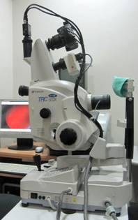 Digital Fundus Fluorescein Angiography (FFA) from Topcon
Fundus Fluorescein Angiography (FFA)
Procedure in which a special dye is injected in the hand and the transit of the dye is captured with a special digital camera and special software. Excellent diagnosis of many retinal conditions like diabetic retinopathy, age related macular degeneration and retinal vascular occlusions can be made.
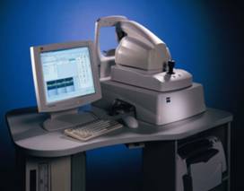 Optical Coherence Tomography( OCT )is a non-invasive technology used for imaging the retina, the multi-layered sensory tissue lining the back of the eye. OCT, the first instrument to allow doctors to see cross-sectional images of the retina, is revolutionizing the early detection and treatment of eye conditions such as macular holes, pre-retinal membranes, macular swelling and even optic nerve damage. There are 10 anatomic layers within the retina. OCT uses the optical backscattering of light to rapidly scan the eye and describe a pixel representation of the anatomic layers within the retina. Each of these ten important layers can be differentiated and their thickness can be measured. For certain conditions, such as age-related macular degeneration and cystoid macular edema, the 45 second OCT procedure is able to reduce or eliminate the need for fluorescein angiography for some patients.
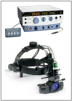 Green laser 532nm
It has all three delivery devices
Viz. Slit lamp, Indirect Ophthalmoscope and endolaser
Laser treatment is done for retinal holes, tears, lattice degeneration, diabetic retinopathy, macular edema and retinal vascular diseases.
Vitrectomy System l is equipped with the most advanced Vitrectomy Systems. It is equipped with 26 G Vitrectomy ensuring spectacular results in Vitreo retinal surgeries.
Zeiss Visu 160 microscope with BIOM
| • Top of the line microscope with XY and motorized zoom |
| • BIOM and SDI for superior viewing of the retina and the vitreous during surgery - The most advanced wide angle viewing system |
| • Panoramic view of the retina with excellent resolution and depth perception during surgery |
| • CCTV with video recording facilities |
All the above state of the art technology ensures excellent results for Vitreoretinal surgeries
© Aggarwal Eye Hospital |
Site designed by Rainbow Web |