 What is Keratoconus?
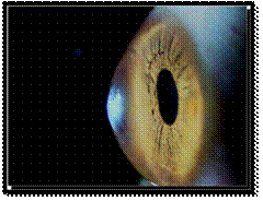
Keratoconus is an uncommon degenerative condition in which the normally round, dome-like cornea (the clear front window of the eye) becomes thin and develops a cone-like bulge.
This abnormal shape prevents the light entering the eye from being focused correctly on the retina and causes distortion of vision.
What causes keratoconus?
Tiny fibers of protein in the eye called collagen help hold the cornea in place and keep it from bulging. When these fibers become weak, they cannot hold the shape and the cornea becomes progressively more cone shaped.
The cause of keratoconus is still not known. It appears to run in families. If you have it and have children, it’s a good idea to have their eyes checked for it starting at age 10. The condition happens more often in people with certain medical problems, including certain allergic conditions. It's possible the condition could be related to chronic eye rubbing.
| • Blurring of vision. |
| • Distortion of vision. |
| • Increased sensitivity to light. |
| • Glare. |
| • Mild eye irritation. |
| • Increased blurring and distortion of your vision |
| • Increased nearsightedness or astigmatism |
| • Frequent change of eyeglass prescription |
| • Inability to wear contact lenses |
Occasionally, keratoconus can advance rapidly, with sudden swelling of the cornea and development of corneal scarring. The swelling occurs when the strain of the cornea's protruding cone-like shape causes a tiny crack to develop. Scar tissue on the cornea causes the cornea to lose its smoothness and clarity. As a result, even more distortion and blurring of vision can occur.
Keratoconus diagnosis
Your ophthalmologist will be able to diagnose keratoconus during a routine eye exam. A slit lamp can be used to diagnose severe cases of keratoconus, but sometimes corneal topography is needed to diagnose the more subtle cases of keratoconus.
| • Spectacles (only beneficial for a limited time in most cases). |
| • Rigid gas permeable contact lenses and other special types of contact lenses. |
| • Corneal collagen cross-linking with Riboflavin. |
| • Intacs. |
| • Toric Phakic IOLs. |
| • Corneal transplant in very advanced cases. |
© Aggarwal Eye Hospital |
Site designed by Rainbow Web |